
Page 124 - Read Online
P. 124
Page 34 of 41 Rao. Vessel Plus 2022;6:25 https://dx.doi.org/10.20517/2574-1209.2021.92
Figure 62. Bar graph illustrating a decrease (P < 0.001) in the left atrial (LA) dimension (A) and LA to aorta (LA/Ao) ratio (B) following
closure patent ductus arteriosus (PDA). Values prior to (Pre), on the day after (Post) occlusion of PDA and at follow-up (FU) were
shown. Note statistically significant reduction (P < 0.01) of LA size and LA:Ao ratio on the day following the procedure; these values
[40]
remain unchanged (P > 0.1) at follow-up. Reproduced from Ref. .
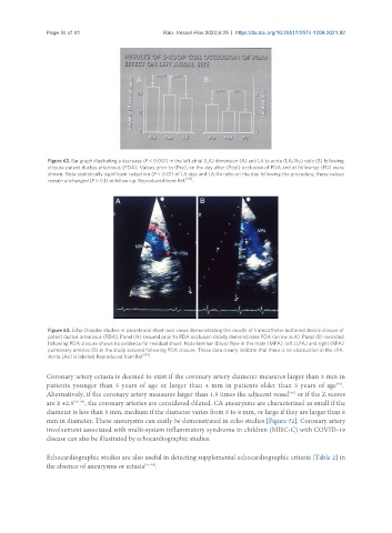
Figure 63. Echo-Doppler studies in parasternal short-axis views demonstrating the results of transcatheter buttoned device closure of
patent ductus arteriosus (PDA). Panel (A) secured prior to PDA occlusion clearly demonstrates PDA (arrow in A). Panel (B) recorded
following PDA closure shows no evidence for residual shunt. Note laminar (blue) flow in the main (MPA), left (LPA) and right (RPA)
pulmonary arteries (B) in the study secured following PDA closure. These data clearly indicate that there is no obstruction in the LPA.
Aorta (Ao) is labeled. Reproduced from Ref. [41] .
Coronary artery ectasia is deemed to exist if the coronary artery diameter measures larger than 3 mm in
[46]
patients younger than 5 years of age or larger than 4 mm in patients older than 5 years of age .
[46]
Alternatively, if the coronary artery measures larger than 1.5 times the adjacent vessel or if the Z scores
are ≥ +2.5 [46-49] , the coronary arteries are considered dilated. CA aneurysms are characterized as small if the
diameter is less than 5 mm, medium if the diameter varies from 5 to 8 mm, or large if they are larger than 8
mm in diameter. These aneurysms can easily be demonstrated in echo studies [Figure 72]. Coronary artery
involvement associated with multi-system inflammatory syndrome in children (MISC-C) with COVID-19
disease can also be illustrated by echocardiographic studies.
Echocardiographic studies are also useful in detecting supplemental echocardiographic criteria [Table 2] in
the absence of aneurysms or ectasia [45-48] .