
Page 126 - Read Online
P. 126
Page 36 of 41 Rao. Vessel Plus 2022;6:25 https://dx.doi.org/10.20517/2574-1209.2021.92
Figure 66. Selected video frames from an apical four-chamber view of a baby with a complete form of atrioventricular septal defect with
severely hypoplastic left ventricle (LV). Echo images in ventricular diastole (A) and systole (B) are shown; the LV hypoplasia is more
obvious in (B). The left atrium (LA), right atrium (RA), and right ventricle (RV) are labeled. Reproduced from Ref. [42] .
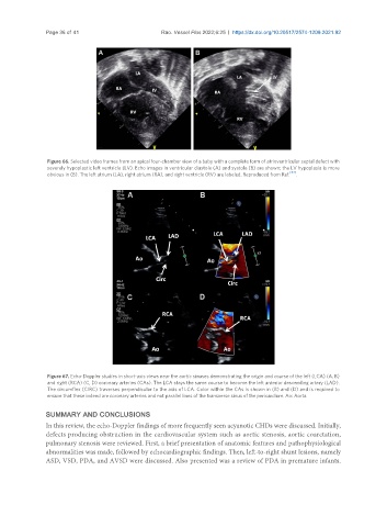
Figure 67. Echo-Doppler studies in short-axis views near the aortic sinuses demonstrating the origin and course of the left (LCA) (A, B)
and right (RCA) (C, D) coronary arteries (CAs). The LCA stays the same course to become the left anterior descending artery (LAD).
The circumflex (CIRC) traverses perpendicular to the axis of LCA. Color within the CAs is shown in (B) and (D) and is required to
ensure that these indeed are coronary arteries and not parallel lines of the transverse sinus of the pericardium. Ao: Aorta.
SUMMARY AND CONCLUSIONS
In this review, the echo-Doppler findings of more frequently seen acyanotic CHDs were discussed. Initially,
defects producing obstruction in the cardiovascular system such as aortic stenosis, aortic coarctation,
pulmonary stenosis were reviewed. First, a brief presentation of anatomic features and pathophysiological
abnormalities was made, followed by echocardiographic findings. Then, left-to-right shunt lesions, namely
ASD, VSD, PDA, and AVSD were discussed. Also presented was a review of PDA in premature infants.