
Page 125 - Read Online
P. 125
Rao. Vessel Plus 2022;6:25 https://dx.doi.org/10.20517/2574-1209.2021.92 Page 35 of 41
Table 2. Supplemental echocardiographic criteria
1. Perivascular brightness
2. Z-scores of left anterior descending or right coronary arteries between 2 and 2.5
3. Lack of vessel tapering
4. Decreased left ventricular function
5. Mitral regurgitation
6. Pericardial effusion
[45]
The presence of 3 or more of the 6 supplemental criteria is considered positive. Reproduced from Ref. .
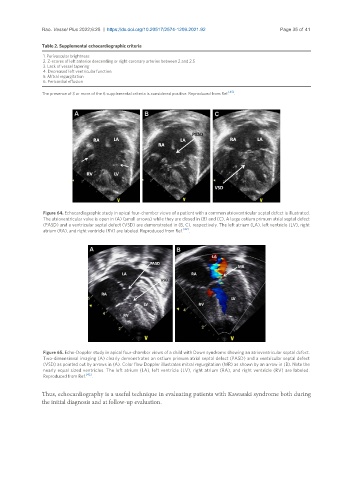
Figure 64. Echocardiographic study in apical four-chamber views of a patient with a common atrioventricular septal defect is illustrated.
The atrioventricular valve is open in (A) (small arrows) while they are closed in (B) and (C). A large ostium primum atrial septal defect
(PASD) and a ventricular septal defect (VSD) are demonstrated in (B, C), respectively. The left atrium (LA), left ventricle (LV), right
atrium (RA), and right ventricle (RV) are labeled. Reproduced from Ref. [42] .
Figure 65. Echo-Doppler study in apical four-chamber views of a child with Down syndrome showing an atrioventricular septal defect.
Two-dimensional imaging (A) clearly demonstrates an ostium primum atrial septal defect (PASD) and a ventricular septal defect
(VSD) as pointed out by arrows in (A). Color flow Doppler illustrates mitral regurgitation (MR) as shown by an arrow in (B). Note the
nearly equal sized ventricles. The left atrium (LA), left ventricle (LV), right atrium (RA), and right ventricle (RV) are labeled.
Reproduced from Ref. [42] .
Thus, echocardiography is a useful technique in evaluating patients with Kawasaki syndrome both during
the initial diagnosis and at follow-up evaluation.