
Page 123 - Read Online
P. 123
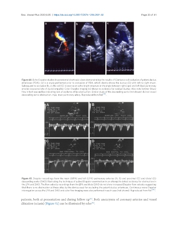
Rao. Vessel Plus 2022;6:25  https://dx.doi.org/10.20517/2574-1209.2021.92       Page 33 of 41


                Figure 60. Echo-Doppler studies in parasternal short-axis views demonstrating the results of Gianturco coil occlusion of patent ductus
                arteriosus (PDA). (a) is a study performed prior to occlusion of PDA, which clearly shows the ductus (D) with left-to-right shunt.
                Subsequent to occlusion (b, c), the coil (C) is seen as an echo-bright structure in the angle between right (rpa) and left (lpa) pulmonary
                arteries (expected site of ductal ampulla). Color Doppler imaging (c) shows no evidence for residual ductus. Also note laminar (blue)
                flow in both rpa and lpa indicating lack of evidence of lpa obstruction. Similar study of the descending aorta (not shown) did not reveal
                descending aortic obstruction. mpa, main pulmonary artery. Reproduced from Ref. [39] .


                Figure 61. Doppler recordings from the main (MPA) and left (LPA) pulmonary arteries (A, B) and proximal (C) and distal (D)
                descending aorta (DAO) illustrating the technique of pulsed Doppler examination in an attempt to detect evidence for obstruction in
                the LPA and DAO. The flow velocity recordings from the LPA and distal DAO do not show increased Doppler flow velocity suggesting
                that there is no obstruction at these sites by the device used for occluding the patent ductus arteriosus. Continuous wave Doppler
                                                                                                       [40]
                interrogation across the LPA and DAO and color flow imaging were also performed in each case (not shown). Reproduced from Ref.  .
               patients, both at presentation and during follow-up . Both aneurysms of coronary arteries and vessel
                                                             [45]
               dilatation (ectasia) [Figure 72] can be illustrated by echo .
                                                              [45]