
Page 128 - Read Online
P. 128
Page 38 of 41 Rao. Vessel Plus 2022;6:25 https://dx.doi.org/10.20517/2574-1209.2021.92
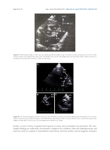
Figure 71. Echo-Doppler study in short-axis view displaying the anomalous origin of the left coronary artery (small arrow) from the
posterior aspect of the pulmonary artery (PA). The division into anterior descending (AD) and circumflex (CIR) coronary arteries are
also shown. Ao: Aorta; ANT: anterior; LT: left; LA: left atrium.
Figure 72. (A) Echocardiographic studies in short-axis view at the level of aortic sinuses illustrating dilated left main (LCA), circumflex
(CIRC) and aneurysmal dilatation (Anu) of the left anterior descending (LAD) coronary arteries (CAs). (B) Diffuse aneurysmal
dilatation of the right CA (RCA Anu). (C) Small aneurysm of the RCA. Ao: Aorta.
Finally, a review of both congenital and acquired coronary artery anomalies was presented. The echo-
Doppler findings are sufficiently characteristic to diagnose the condition, define the pathophysiology, and
assess the need for surgical or transcatheter intervention such that studies such as magnetic resonance