
Page 127 - Read Online
P. 127
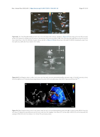
Rao. Vessel Plus 2022;6:25  https://dx.doi.org/10.20517/2574-1209.2021.92       Page 37 of 41


                Figure 68. (A) Echo-Doppler study in short-axis view near the aortic sinuses, similar to  [Figure 67], focusing on to the left coronary
                artery (CA) shows its division into left anterior descending (AD) and circumflex (CIR) CAs. Note the red-coded flow in the AD and the
                blue-coded flow in the CIR. (B) Echo-Doppler study, similar to [Figure 67A], but focusing on the right CA (RCA) showing its origin from
                the right sinus with color flow inside it. AO: Aorta.


                Figure 69. Echo-Doppler study in short-axis view near the aortic sinuses demonstrating the aberrant origin of the left coronary artery
                                                                                             [43]
                from the right sinus of Valsalva, traversing between the aorta (AO) and pulmonary artery (PA). Reproduced from Ref.  .


                Figure 70. Echo-Doppler study in short-axis views near the aortic sinuses demonstrating aberrant right coronary artery (RCA) from the
                left sinus of Valsalva by two-dimensional (A) and color flow (B) imaging. Color flow (CF) at the origin of RCA (B) and intramural (IM)
                passage of the RCA (A, B) are shown. Ao: Aorta; PA: pulmonary artery.