
Page 119 - Read Online
P. 119
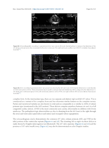
Rao. Vessel Plus 2022;6:25  https://dx.doi.org/10.20517/2574-1209.2021.92       Page 29 of 41


                Figure 52. Echocardiographic recording in a parasternal short-axis view by M-mode illustrating how to measure the diameters of the
                left atrium (LA) and aorta (Ao). Increased LA/Ao ratio is observed. The right ventricular outflow tract (RVOT) is labeled. Reproduced
                from Ref. [36] .


                Figure 53. Echo recording in parasternal short-axis projection showing the left ventricular (LV) internal dimensions in end-diastole
                (LVIDd) and systole (LVIDs). These dimensions may be used to compute fractional shortening (%FS), along with other data shown in
                the insert. Inter-ventricular septum (IVS), left ventricular posterior wall (LVPW), and right ventricle (RV) are also labeled. Reproduced
                from Ref. [36] .


               complete form. In the intermediate type, there are two separate and distinct right and left AV valves. This is
               considered as a variant of the complete form and has otherwise similar features as the complete variety.
               Partial and transitional varieties are also known to exist and are comparable to or similar to ASDs of ostium
               primum type (mentioned in the ASD section). These defects comprise anywhere between 4% and 5% of all
               congenital cardiac defects. AVSD is the most commonly seen cardiac abnormality in children with Down
               syndrome. The pathophysiologic abnormalities are due to shunting from the left heart to the right through
               the atrial and ventricular septal defects and mitral (and tricuspid) valvar regurgitation.

               The echocardiogram clearly demonstrates the common AV valve, ostium primum ASD, and VSD in the
               inlet portion of the ventricular septum [Figures 64 and 65]. The shunting left-to-right via these defects is
               easily shown by Doppler interrogation (not illustrated). The AV valve anatomy [Figures 64 and 65] and the
               presence of AV valve insufficiency [Figure 65] may also be demonstrated on echo-Doppler studies.