
Page 117 - Read Online
P. 117
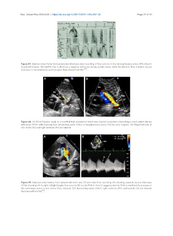
Rao. Vessel Plus 2022;6:25  https://dx.doi.org/10.20517/2574-1209.2021.92       Page 27 of 41


                Figure 47. Selected video frame from parasternal short-axis view recording of flow velocity in the main pulmonary artery (PA) (insert)
                by pulsed Doppler. Normal PA flow is shown as a negative deflection during systole phase, while the diastolic flow of patent ductus
                                                           [35]
                arteriosus is represented by positive signal. Reproduced from Ref.  .


                Figure 48. (A) Echo-Doppler study in a modified high parasternal short-axis (ductal) projection, illustrating a small patent ductus
                arteriosus (PDA) with shunting from descending aorta (DAo) to the pulmonary artery (PA) by color Doppler. (B) Magnified view of
                (A). Aorta (Ao) and right ventricle (RV) are labeled.


                Figure 49. Selected video frames from parasternal short-axis 2D and color flow recording (A) showing a patent ductus arteriosus
                (PDA) shunting left-to-right. A high Doppler flow velocity (B) via the PDA (≈ 4 m/s) suggests that the PDA is small and the pressure in
                the pulmonary artery is low. Aorta (Ao), diastolic (D), descending aorta (DAo), right ventricle (RV), and systolic (S) are labeled.
                Reproduced from Ref. [16] .