
Page 118 - Read Online
P. 118
Page 28 of 41 Rao. Vessel Plus 2022;6:25 https://dx.doi.org/10.20517/2574-1209.2021.92
Figure 50. Echo images from apical four-chamber projections illustrating a dilated left atrium (LA) and left ventricle (LV). The images
are recorded with open (MVO) and closed (MVC) mitral valve leaflets respectively, depicted in (A) and (B). However, this appearance
of dilated LA and LV is very subjective. The right atrium (RA) and right ventricle (RV) are marked. Reproduced from Ref. [36] .
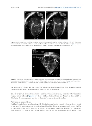
Figure 51. Echo images from an apical four-chamber projection illustrating plotting of the area of the left atrium (LA). While the area
measurements can be made (see insert at the top left of the image), normal values for several premature baby groups have not been
established. The left ventricle (LV), right atrium (RA), and right ventricle (RV) are labeled. Reproduced from Ref. [36] .
anterograde DAo diastolic flow is not observed. In babies with medium and large PDAs in association with
compromised respiratory status, a diagnosis of hsPDA may be established [36-38] .
Echocardiographic examination has also been found valuable in assessing outcomes following either
surgical or transcatheter [Figures 60-63] closure of PDAs. Residual shunts and obstruction of the left PA or
DAo by the device components may also be illustrated by echocardiographic studies.
Atrioventricular septal defect
Atrial and ventricular septal defects along with clefts in the mitral and/or tricuspid valves, previously named
as common AV canal or complete form of endocardial cushion defect, are now commonly termed AVSDs.
In the complete type, a VSD is present in the inlet portion of the ventricular septum; this VSD adjoins
(contiguous with) a primum ASD. A common AV valve with a solitary valve annulus is present in the