
Page 17 - Read Online
P. 17
Castán et al. Radiology of hepatocarcinoma in non-cirrhotic patients
A B
C D
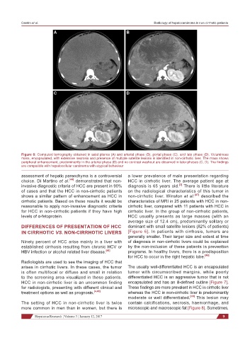
Figure 6: Computed tomography obtained in axial planes (A) and arterial phase (B), portal phase (C), and late phase (D). Voluminous
mass, encapsulated, with extensive necrosis and presence of multiple satellite lesions is identified in non-cirrhotic liver. The mass shows
peripheral enhancement, predominantly in the arterial phase (B) and no contrast washout are observed in later phases (C, D). The findings
are compatible with hepatocellular carcinoma with atypical behaviour
assessment of hepatic parenchyma is a controversial a lower prevalence of male presentation regarding
choice. Di Martino et al. [44] demonstrated that non- HCC in cirrhotic liver. The average patient age at
[3]
invasive diagnostic criteria of HCC are present in 90% diagnosis is 65 years old. There is little literature
of cases and that the HCC in non-cirrhotic patients on the radiological characteristics of this tumor in
shows a similar pattern of enhancement as HCC in non-cirrhotic liver. Winston et al. [47] described the
cirrhotic patients. Based on these results it would be characteristics of MRI in 25 patients with HCC in non-
reasonable to apply non-invasive diagnostic criteria cirrhotic liver, compared with 11 patients with HCC in
for HCC in non-cirrhotic patients if they have high cirrhotic liver. In the group of non-cirrhotic patients,
levels of α-fetoprotein. HCC usually presents as large masses (with an
average size of 12.4 cm), predominantly solitary or
DIFFERENCES OF PRESENTATION OF HCC dominant with small satellite lesions (82% of patients)
IN CIRRHOTIC VS. NON-CIRRHOTIC LIVERS [Figure 6]. In patients with cirrhosis, tumors are
generally smaller. Their larger size and extent at time
Ninety percent of HCC arise mainly in a liver with of diagnosis in non-cirrhotic livers could be explained
established cirrhosis resulting from chronic HCV or by the non-inclusion of these patients in prevention
HBV infection or alcohol related liver disease. [45] programs. In healthy livers, there is a predisposition
for HCC to occur in the right hepatic lobe. [48]
Radiologists are used to see the imaging of HCC that
arises in cirrhotic livers. In these cases, the tumor The usually well-differentiated HCC is an encapsulated
is often multifocal or diffuse and small in relation tumor with circumscribed margins, while poorly
to the screening area visualized in these patients. differentiated HCC is an aggressive tumor that is not
HCC in non-cirrhotic liver is an uncommon finding encapsulated and has an ill-defined outline [Figure 7].
for radiologists, presenting with different clinical and These findings are more prevalent in HCC in cirrhotic liver
treatment options as well as prognosis. [4,46] whereas the HCC in non-cirrhotic liver is predominantly
moderate or well differentiated. [49] This lesion may
The setting of HCC in non-cirrhotic liver is twice contain calcifications, necrosis, haemorrhage, and
more common in men than in women, but there is microscopic and macroscopic fat [Figure 8]. Sometimes,
Hepatoma Research ¦ Volume 3 ¦ January 12, 2017 9