
Page 19 - Read Online
P. 19
Castán et al. Radiology of hepatocarcinoma in non-cirrhotic patients
A B
C D
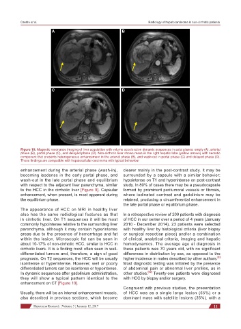
Figure 10: Magnetic resonance imaging of liver acquisition with volume acceleration dynamic sequences in axial planes: empty (A), arterial
phase (B), portal phase (C), and delayed phase (D). Non-cirrhotic liver shows mass in the right hepatic lobe (yellow arrows) with necrotic
component that presents heterogeneous enhancement in the arterial phase (B), and wash-out in portal phase (C) and delayed phase (D).
These findings are compatible with hepatocellular carcinoma with typical behaviour
enhancement during the arterial phase (wash-in), clearer mainly in the post-contrast study. It may be
becoming isodense in the early portal phase, and surrounded by a capsule with a similar behavior:
wash-out in the late portal phase and equilibrium hypointense on T1 and hyperintense on post-contrast
with respect to the adjacent liver parenchyma, similar study. In 80% of cases there may be a pseudocapsule
to the HCC in the cirrhotic liver [Figure 9]. Capsular formed by prominent peritumoral vessels or fibrosis,
enhancement, when present, is most apparent during where iodinated contrast and gadolinium may be
the equilibrium phase. retained, producing a circumferential enhancement in
the late portal phase or equilibrium phase.
The appearance of HCC on MRI in healthy liver
also has the same radiological features as that In a retrospective review of 209 patients with diagnosis
in cirrhotic liver. On T1 sequences it will be most of HCC in our center over a period of 4 years (January
commonly hypointense relative to the surrounding liver 2010 - December 2014), 23 patients were selected
parenchyma, although it may contain hyperintense with healthy liver by histological criteria (liver biopsy
areas due to the presence of hemorrhage and fat or surgical resection piece) and/or a combination
within the lesion. Microscopic fat can be seen in of clinical, analytical criteria, imaging and hepatic
about 10-17% of non-cirrhotic HCC, similar to HCC in hemodynamics. The average age at diagnosis in
cirrhotic livers. It is a finding most often seen in well- these patients was 70 years old, with no significant
differentiated tumors and, therefore, a sign of good differences in distribution by sex, as opposed to the
[3]
prognosis. On T2 sequences, the HCC will be usually higher incidence in males described by other authors.
isointense or hyperintense. However, well or poorly Most diagnostic testing was initiated by the presence
differentiated tumors can be isointense or hypointense. of abdominal pain or abnormal liver profiles, as in
In dynamic sequences after gadolinium administration, other studies. [50] Twenty-one patients were diagnosed
they will show a typical pattern identical to the with HCC by biopsy and/or surgery.
enhancement on CT [Figure 10].
Congruent with previous studies, the presentation
Usually, there will be an internal enhancement mosaic, of HCC was as a single large lesion (65%) or a
also described in previous sections, which become dominant mass with satellite lesions (35%), with a
Hepatoma Research ¦ Volume 3 ¦ January 12, 2017 11