
Page 23 - Read Online
P. 23
Castán et al. Radiology of hepatocarcinoma in non-cirrhotic patients
A B C
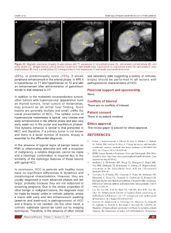
Figure 15: Magnetic resonance imaging in axial planes with T1 sequences in noncontrast phase (A), and arterial contrast phase (B), and
portal phase (C). Multiple lesions (yellow arrows) scattered in right hepatic lobe, hypointense in noncontrast phase (A) and uptake in early
arterial phase (B), held in portal phase (C) corresponding to liver metastases of pancreatic neuroendocrine tumor
(25%), or predominantly cystic (15%). It shows and laboratory data suggesting a history of cirrhosis,
peripheral enhancement in the arterial phase. In MRI it biopsy should be performed in all lesions with
is hypointense on T1 and hyperintense on T2 and with pathognomonic characteristics of HCC.
an enhancement after administration of gadolinium
similar to that obtained in CT. Financial support and sponsorship
None.
In addition to the metastatic neuroendocrine tumors,
other tumors with hypervascular appearance such Conflicts of interest
as thyroid tumors, renal tumors or melanomas, There are no conflicts of interest.
may present as an initial liver finding. Such
lesions are generally multiple and small, unlike the Patient consent
usual presentation of HCC. The uptake curve of
hypervascular metastases is typical: very intense and There is no patient involved.
early enhancement in the arterial phase and also very
early wash-out in the portal and equilibrium phases. Ethics approval
This dynamic behavior is similar to that presented in This review paper is waived for ethics approval.
HCC and therefore, if a primary tumor is not known
and there is a small number of lesions, biopsy is REFERENCES
essential for the differential diagnosis.
1. Ferlay J, Soerjomataram I, Dikshit R, Eser S, Mathers C, Rebelo
In the absence of typical signs of benign lesion as M, Parkin DM, Forman D, Bray F. Cancer incidence and mortality
FNH or inflammatory adenoma and with a suspicion worldwide: sources, methods and major patterns in GLOBOCAN
2012. Int J Cancer 2015;136:E359-86.
of malignancy, a reliable diagnosis cannot be made 2. SEER Cancer Statistics Factsheets: Liver and Intrahepatic Bile Duct.
and a histologic confirmation is required due to the Available from: http://seer.cancer.gov/statfacts/html/livibd.html. [Last
similarity of the radiologic features of these lesions accessed on Aug 29 2016].
with typical HCC. 3. Gaddikeri S, McNeeley MF, Wang CL, Bhargava P, Dighe MK,
Yeh MM, Dubinsky TJ, Kolokythas O, Lalwani N. Hepatocellular
In conclusion, HCC in patients with healthy livers carcinoma in the noncirrhotic liver. AJR Am J Roentgenol
have no significant differences in dynamics and 4. 2014;203:W34-47.
Trevisani F, D’Intino PE, Caraceni P, Pizzo M, Stefanini GF,
morphological characteristics. However, they are Mazziotti A, Grazi GL, Gozzetti G, Gasbarrini G, Bernardi M.
usually diagnosed in more advanced phases and are Etiologic factors and clinical presentation of hepatocellular carcinoma.
larger, probably because they are not subjected to Differences between cirrhotic and noncirrhotic Italian patients. Cancer
screening programs. Due to the similar properties of 1995;75:2220-32.
other benign or malignant lesions, the diagnosis must 5. Lee JH, Lee JM, Kim SJ, Baek JH, Yun SH, Kim KW, Han JK,
be made by biopsy unlike in cirrhotic patients, where Choi BI. Enhancement patterns of hepatocellular carcinomas on
a lesion with early and late enhancement washing multiphasicmultidetector row CT: comparison with pathological
differentiation. Br J Radiol 2012;85:e573-83.
(wash-in and wash-out) is pathognomonic of HCC 6. Iavarone M, Sangiovanni A, Forzenigo LV, Massironi S, Fraquelli
and a biopsy is not needed. On the other hand, a M, Aghemo A, Ronchi G, Biondetti P, Roncalli M, Colombo M.
cirrhotic substrate cannot be ruled out by imaging Diagnosis of hepatocellular carcinoma in cirrhosis by dynamic contrast
techniques. Therefore, in the absence of other clinical imaging: the importance of tumor cell differentiation. Hepatology
Hepatoma Research ¦ Volume 3 ¦ January 12, 2017 15