
Page 12 - Read Online
P. 12
Castán et al. Radiology of hepatocarcinoma in non-cirrhotic patients
A vascular pattern in HCC, more frequent in those that
are moderately differentiated [11] [Figure 4]. Contrast
agent flows exclusively through the intravascular
space, without passing to the interstitial liquid, thus
explaining some differences with the typical features
found in CT or MRI. However, other reports have not
found significant differences. Wilson et al. [12] reported
no differences in the dynamic behavior among
CEUS, MRI and CT. Giorgio et al. [13] did not find any
difference between CEUS and CT. Nevertheless,
Liu et al. [14] reported different results for small
lesions detected by CEUS and CT. In their report, a
B good correlation was found between both imaging
techniques among lesions greater than 2 cm, but
there was a low correlation among lesions measuring
1-2 cm. Possible explanations for this discrepancy are
the different distribution of contrast agents, the various
thickness of the slices of CT, and the effect of the
direct time changes measured with CEUS. A cirrhotic
background may also cause atypical patterns due to
the progressive arterialization of the small lesions.
These results suggest that more research is needed
to determine the usefulness of CEUS in the diagnosis
of HCC.
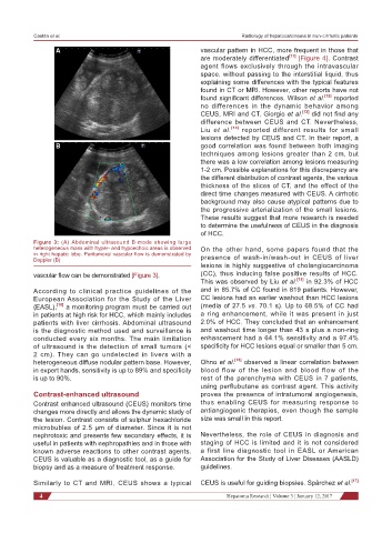
Figure 3: (A) Abdominal ultrasound B-mode showing large
heterogeneous mass with hyper- and hypoechoic areas is observed On the other hand, some papers found that the
in right hepatic lobe. Peritumoral vascular flow is demonstrated by
Doppler (B) presence of wash-in/wash-out in CEUS of liver
lesions is highly suggestive of cholangiocarcinoma
vascular flow can be demonstrated [Figure 3]. (CC), thus inducing false positive results of HCC.
This was observed by Liu et al. [15] in 92.3% of HCC
According to clinical practice guidelines of the and in 85.7% of CC found in 819 patients. However,
European Association for the Study of the Liver CC lesions had an earlier washout than HCC lesions
(EASL), [10] a monitoring program must be carried out (media of 27.5 vs. 70.1 s). Up to 68.5% of CC had
in patients at high risk for HCC, which mainly includes a ring enhancement, while it was present in just
patients with liver cirrhosis. Abdominal ultrasound 2.0% of HCC. They concluded that an enhancement
is the diagnostic method used and surveillance is and washout time longer than 43 s plus a non-ring
conducted every six months. The main limitation enhancement had a 64.1% sensitivity and a 97.4%
of ultrasound is the detection of small tumors (< specificity for HCC lesions equal or smaller than 5 cm.
2 cm). They can go undetected in livers with a
heterogeneous diffuse nodular pattern base. However, Ohno et al. [16] observed a linear correlation between
in expert hands, sensitivity is up to 89% and specificity blood flow of the lesion and blood flow of the
is up to 90%. rest of the parenchyma with CEUS in 7 patients,
using perflubutane as contrast agent. This activity
Contrast-enhanced ultrasound proves the presence of intratumoral angiogenesis,
Contrast enhanced ultrasound (CEUS) monitors time thus enabling CEUS for measuring response to
changes more directly and allows the dynamic study of antiangiogenic therapies, even though the sample
the lesion. Contrast consists of sulphur hexachloride size was small in this report.
microbubles of 2.5 μm of diameter. Since it is not
nephrotoxic and presents few secondary effects, it is Nevertheless, the role of CEUS in diagnosis and
useful in patients with nephropathies and in those with staging of HCC is limited and it is not considered
known adverse reactions to other contrast agents. a first line diagnostic tool in EASL or American
CEUS is valuable as a diagnostic tool, as a guide for Association for the Study of Liver Diseases (AASLD)
biopsy and as a measure of treatment response. guidelines.
Similarly to CT and MRI, CEUS shows a typical CEUS is useful for guiding biopsies. Spârchez et al. [17]
4 Hepatoma Research ¦ Volume 3 ¦ January 12, 2017