
Page 13 - Read Online
P. 13
Castán et al. Radiology of hepatocarcinoma in non-cirrhotic patients
A B
C D E
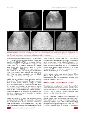
Figure 4: Abdominal ultrasound (A) and with contrast at 23 s (B), 30 s (C), 1 min (D), and 5 min (E). Hypoechoic lesion in right hepatic lobe
corresponding to hepatocellular carcinoma with a typical vascular pattern: early uptake in arterial phases (B, C), isoechogenic respect to
surrounded liver parenchyma in portal phase (D) and wash-out contrast in late phase (E)
prospectively compared conventional US and CEUS lower rates in patients with tumor recurrence
in 171 cirrhotic and non-cirrhotic patients. Biopsy was compared with those without recurrence. On the other
possible with CEUS in 97.6% of the cases, obtaining hand, the expression levels of basic fibroblast growth
one sole sample in 43.0% of them, compared with factor in the recurrence group were higher than those
23.4% using US. In general, sensitivity was greater in the non-recurrence group. Xia et al. [20] and other
with CEUS (96.5% vs. 81.5%), also in cirrhotic reports have shown a greater sensitivity of CEUS
patients (95.2% vs. 75.0%), in lesions greater than 6 compared to CT when detecting residual tumor after
cm (97.8% vs. 82.0%) and in poorly visualized lesions chemoembolization (58.1% vs. 39.5%).
(100.0% vs. 66.6%). When histology was inconclusive
with US a new biopsy was performed with CEUS, CEUS has not shown better sensitivity than CT or
obtaining a final diagnosis in every case. MRI when looking for late recurrence. Thus, these two
techniques are the gold standard for the long term
CEUS may be useful also to monitor tumor response follow-up of patients with HCC.
to treatment. With antiangiogenic therapy, changes in
tumoral vascularization precede changes in tumoral MANAGEMENT IN DIAGNOSIS OF HCC
size. A complete response may be considered when
there is no enhancement at any time. Irregular The objective is early detection. In early stages, radical
enhancement and/or eccentrical or peripheral nodules treatment and improved prognosis are possible. The
suggest the presence of residual tumor. [18] Using a pathological diagnosis of the tumor involves biopsy of
quantitative analysis an individualized treatment could the lesion. It is an invasive technique including risks
be done, but more research is needed to establish such as bleeding or tumor seeding.
this indication for CEUS.
In 2001, diagnostic criteria for the management of
CEUS performed 60 min after radiofrequency ablation nodular lesions in the cirrhotic liver were established.
or alcoholization of HCC may monitor the efficacy of These criteria favor an early non-invasive diagnosis,
the treatment. [18] Gao et al. [19] measured the different preventing biopsy in some cases. In the latest update
peak enhancement of contrast between tumor and of the clinical practice guidelines of the EASL (2012), [10]
surrounding parenchyma and encountered significantly the criteria are as follows: (1) nodules > 2 cm can be
Hepatoma Research ¦ Volume 3 ¦ January 12, 2017 5