
Page 10 - Read Online
P. 10
Castán et al. Radiology of hepatocarcinoma in non-cirrhotic patients
in underdeveloped countries. In developed countries, later stages. Sometimes, the edges are imprecise,
most HCC originate in a setting of alcoholic cirrhosis which also determines more aggressive tumors.
or non-alcoholic steatosis related to obesity. However, Growth is usually expansive although there may
there is an incidence of 0.5-1% per year in patients be transcapsular infiltration into the surrounding
[2]
with non-cirrhotic livers. Usually, such patients are parenchyma.
not subject to monitoring prevention programs and
so HCC detection is usually late and secondary to However, a high percentage of patients do not
symptoms produced by the tumor. Less frequent demonstrate pathognomonic HCC criteria, showing
risk factors are type II diabetes and metabolic atypical features. Thus, in a retrospective study
[5]
syndrome, congenital diseases such as hereditary of 243 patients conducted by Lee et al., the
hemochromatosis, tobacco, parasitic infections or most typical behavior of tumors corresponded to
genotoxin intake. The average age at diagnosis of moderately differentiated HCC. A high percentage
HCC is 63 years old, with an incidence three times of cases showed atypical behavior (43.6%). Most
higher in men than in women. [2] of these tumors corresponded histologically to well
Clinically, it is a silent disease in early stages. When A
symptoms appear, the most common is abdominal pain
[3]
(52%). Less common symptoms are chronic diarrhea,
jaundice, fever, or paraneoplastic syndromes such
as hypercalcemia or hypoglycemia. It may occur with
increased serum levels of alpha-fetoprotein, considered
[4]
indicative of HCC above 400 ng/dL. However, this
determination has low sensitivity and specificity for
diagnosis and for monitoring.
RADIOLOGICAL DIAGNOSIS OF HCC
There are three basic diagnostic tests: computed B
tomography (CT), magnetic resonance imaging (MRI)
and ultrasound (US).
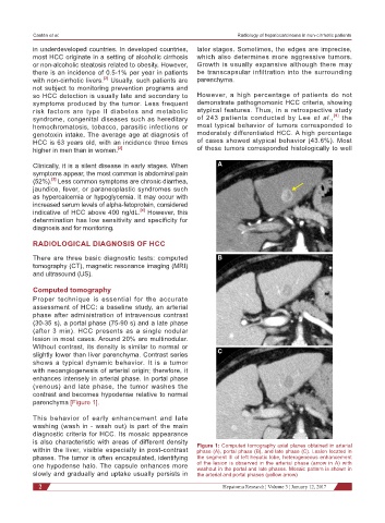
Computed tomography
Proper technique is essential for the accurate
assessment of HCC: a baseline study, an arterial
phase after administration of intravenous contrast
(30-35 s), a portal phase (75-90 s) and a late phase
(after 3 min). HCC presents as a single nodular
lesion in most cases. Around 20% are multinodular.
Without contrast, its density is similar to normal or
slightly lower than liver parenchyma. Contrast series C
shows a typical dynamic behavior. It is a tumor
with neoangiogenesis of arterial origin; therefore, it
enhances intensely in arterial phase. In portal phase
(venous) and late phase, the tumor washes the
contrast and becomes hypodense relative to normal
parenchyma [Figure 1].
This behavior of early enhancement and late
washing (wash in - wash out) is part of the main
diagnostic criteria for HCC. Its mosaic appearance
is also characteristic with areas of different density Figure 1: Computed tomography axial planes obtained in arterial
within the liver, visible especially in post-contrast phase (A), portal phase (B), and late phase (C). Lesion located in
phases. The tumor is often encapsulated, identifying the segment III of left hepatic lobe, heterogeneous enhancement
one hypodense halo. The capsule enhances more of the lesion is observed in the arterial phase (arrow in A) with
washout in the portal and late phases. Mosaic pattern is shown in
slowly and gradually and uptake usually persists in the arterial and portal phases (yellow arrow)
2 Hepatoma Research ¦ Volume 3 ¦ January 12, 2017