
Page 98 - Read Online
P. 98
Page 8 of 41 Rao. Vessel Plus 2022;6:25 https://dx.doi.org/10.20517/2574-1209.2021.92
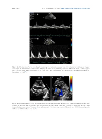
Figure 10. Selected video frames from Doppler recordings from subcostal views of two different patients. In (A), pulse Doppler
recording of the abdominal aorta demonstrates damping of the flow velocity indicating coarctation of the aorta. In (B), a similar Doppler
recording of a normal child illustrates a pulsatile signal with a high magnitude and arterial variety of flow signal with a sharp rise.
Reproduced from Ref. [10] .
Figure 11. Echocardiographic study in suprasternal notch view imaging the arch of the aorta (Ao) by two-dimensional (A) and color
Doppler (B) recording of a newborn baby who was diagnosed to have tricuspid atresia with transposition of the great arteries. These
images illustrate coarctation of the aorta (CoA) and hypoplasia of the transverse portion of the aortic arch (TAA). Descending aorta
(DAo) is labeled. Reproduced from Ref. [10] .