
Page 95 - Read Online
P. 95
Rao. Vessel Plus 2022;6:25 https://dx.doi.org/10.20517/2574-1209.2021.92 Page 5 of 41
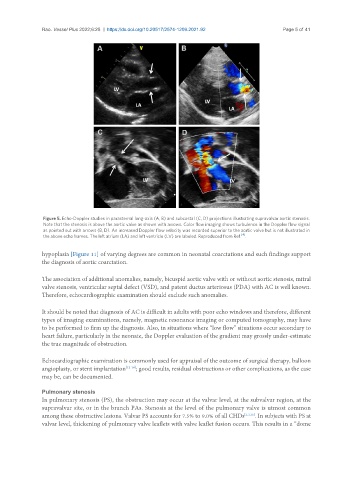
Figure 5. Echo-Doppler studies in parasternal long-axis (A, B) and subcostal (C, D) projections illustrating supravalvar aortic stenosis.
Note that the stenosis is above the aortic valve as shown with arrows. Color flow imaging shows turbulence in the Doppler flow signal
as pointed out with arrows (B, D). An increased Doppler flow velocity was recorded superior to the aortic valve but is not illustrated in
[4]
the above echo frames. The left atrium (LA) and left ventricle (LV) are labeled. Reproduced from Ref. .
hypoplasia [Figure 11] of varying degrees are common in neonatal coarctations and such findings support
the diagnosis of aortic coarctation.
The association of additional anomalies, namely, bicuspid aortic valve with or without aortic stenosis, mitral
valve stenosis, ventricular septal defect (VSD), and patent ductus arteriosus (PDA) with AC is well known.
Therefore, echocardiographic examination should exclude such anomalies.
It should be noted that diagnosis of AC is difficult in adults with poor echo windows and therefore, different
types of imaging examinations, namely, magnetic resonance imaging or computed tomography, may have
to be performed to firm up the diagnosis. Also, in situations where “low flow” situations occur secondary to
heart failure, particularly in the neonate, the Doppler evaluation of the gradient may grossly under-estimate
the true magnitude of obstruction.
Echocardiographic examination is commonly used for appraisal of the outcome of surgical therapy, balloon
angioplasty, or stent implantation [12-14] ; good results, residual obstructions or other complications, as the case
may be, can be documented.
Pulmonary stenosis
In pulmonary stenosis (PS), the obstruction may occur at the valvar level, at the subvalvar region, at the
supravalvar site, or in the branch PAs. Stenosis at the level of the pulmonary valve is utmost common
among these obstructive lesions. Valvar PS accounts for 7.5% to 9.0% of all CHDs [1,3,15] . In subjects with PS at
valvar level, thickening of pulmonary valve leaflets with valve leaflet fusion occurs. This results in a “dome