
Page 93 - Read Online
P. 93
Rao. Vessel Plus 2022;6:25 https://dx.doi.org/10.20517/2574-1209.2021.92 Page 3 of 41
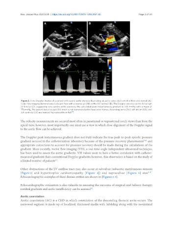
Figure 2. Echo-Doppler studies of a patient with severe aortic stenosis illustrating an aortic valve (AV) which is thick and domed (A).
Color flow imaging demonstrates turbulent flow with a narrow jet (NJ) at the AV (arrow) (B). The Doppler velocity via the AV is high
(> 6 m/s) (C), suggesting very severe aortic stenosis; the calculated peak instantaneous gradient is 148 mmHg with a mean of
75 mmHg. The patient has a bicuspid AV which is not demonstrated in these echo frames. Ascending aorta (Ao), left atrium (LA), and
[4]
left ventricle (LV) are marked. Reproduced from Ref. .
The velocity measurements are secured most often in parasternal or suprasternal notch views than from the
apical view; however, most importantly one must use a view in which close alignment of the Doppler signal
to the aortic flow can be achieved.
The Doppler peak instantaneous gradient does not truly indicate the true peak-to-peak systolic pressure
gradient secured in the catheterization laboratory because of the pressure recovery phenomenon and
[5,6]
appropriate corrections to account for pressure recovery should be made during the calculations of the
gradient. More recently, vector flow imaging (VFI), a real-time angle-independent ultrasound technique,
has been used to assess the aortic gradients. VFI values seem to have a better correlation with catheter-
measured gradients than conventional Doppler gradients; however, this observation is based on the study of
[7]
a limited number of patients .
Other obstructions of the LV outflow tract may also occur at subvalvar (subaortic membranous stenosis
[Figure 3] and hypertrophic cardiomyopathy [Figure 4]) and supravalvar [Figure 5] sites .
[1-3]
Echocardiographic examples of these disease entities are shown in [Figures 3-5].
Echocardiographic evaluation is also valuable in assessing the outcome of surgical and balloon therapy;
residual gradients and aortic insufficiency can be assessed .
[8]
Aortic coarctation
Aortic coarctation (AC) is a CHD in which constriction of the descending thoracic aorta occurs. The
narrowed segment is made up of localized, thickened media with infolding along with the neointimal