
Page 97 - Read Online
P. 97
Rao. Vessel Plus 2022;6:25 https://dx.doi.org/10.20517/2574-1209.2021.92 Page 7 of 41
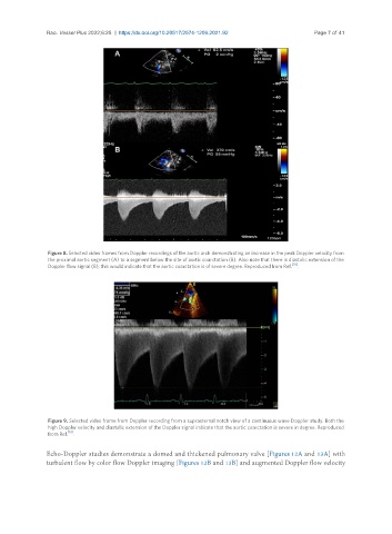
Figure 8. Selected video frames from Doppler recordings of the aortic arch demonstrating an increase in the peak Doppler velocity from
the proximal aortic segment (A) to a segment below the site of aortic coarctation (B). Also note that there is diastolic extension of the
Doppler flow signal (B); this would indicate that the aortic coarctation is of severe degree. Reproduced from Ref. [10] .
Figure 9. Selected video frame from Doppler recording from a suprasternal notch view of a continuous wave Doppler study. Both the
high Doppler velocity and diastolic extension of the Doppler signal indicate that the aortic coarctation is severe in degree. Reproduced
from Ref. [10] .
Echo-Doppler studies demonstrate a domed and thickened pulmonary valve [Figures 12A and 13A] with
turbulent flow by color flow Doppler imaging [Figures 12B and 13B] and augmented Doppler flow velocity