
Page 101 - Read Online
P. 101
Rao. Vessel Plus 2022;6:25 https://dx.doi.org/10.20517/2574-1209.2021.92 Page 11 of 41
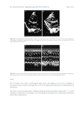
Figure 14. Two-dimensional echocardiographic studies in parasternal long-axis view before (A) and after (B) balloon pulmonary
valvuloplasty, illustrating a reduction in the size of the right ventricle (RV). Aorta (Ao), left atrium (LA), and left ventricle (LV) are
labeled. Reproduced from Ref. [17] .
Figure 15. M-mode echocardiographic tracings in parasternal long-axis view before (A) and after (B) balloon pulmonary valvuloplasty,
illustrating a reduction in the size of the right ventricle (RV). The left ventricle (LV) is labeled. Reproduced from Ref. [17] .
defect.
The 2D study is also useful in defining the atrial septal rims [Figures 25 and 26] for suitability of
transcatheter device occlusion, although TEE or ICE in the catheterization laboratory is usually utilized for
this purpose.
Since there is an increased prevalence of deficient atrial septal rims, particularly in large ASDs [28,29] , a careful
evaluation of septal rims by TEE [Figure 27] or ICE should be performed in the catheterization laboratory
prior to device implantation.