
Page 106 - Read Online
P. 106
Page 16 of 41 Rao. Vessel Plus 2022;6:25 https://dx.doi.org/10.20517/2574-1209.2021.92
Figure 25. Two-dimensional echocardiograms of an atrial septal defect (ASD) to illustrate the atrial septal rims in apical four-chamber
(A), subcostal long-axis (B), parasternal short-axis (C), and subcostal short-axis (D) views. The unfilled arrows point to the ASD.
Adequate-sized septal rims are seen in each image. This patient is deemed to be an appropriate patient for percutaneous occlusion.
Reproduced from Ref. [26] . Ao: Aorta; LA: left atrium; LV: left ventricle; RA: right atrium; RV: right ventricle.
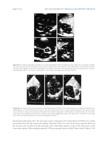
Figure 26. Two-dimensional echocardiograms of atrial septal defects (ASDs) in three different patients to illustrate deficient atrial
septal margins. In (A) both the inferior and superior rims are small, in (B) the inferior margin is deficient, and in (C) the superior margin
is tiny; the rims are shown with arrowheads. These ASDs are considered unsuitable for transcatheter occlusion because the rims of the
atrial septum are inadequate for the device to achieve a good grasp on the atrial septum. Left atrium (LA), left ventricle (LV), right
atrium (RA), and right ventricle (RV) are marked. Reproduced from Ref. [27] .
beneath the pulmonary valve. The next type is atrio-ventricular (AV) septal defects (AVSDs) (8%), which
are positioned in the inlet ventricular septum. Muscular VSDs (5%-20%) are the most common defects in
the neonate and are placed in the muscular inter-ventricular septum, some in the apical part of the
ventricular septum. When multiple muscular VSDs are present, they are called “Swiss-cheese” defects. VSD