
Page 111 - Read Online
P. 111
Rao. Vessel Plus 2022;6:25 https://dx.doi.org/10.20517/2574-1209.2021.92 Page 21 of 41
Figure 35. Echo-Doppler studies in modified subcostal four-chamber projections demonstrating an ostium primum atrial septal defect
(PASD) (A) with shunting from the left (LA) to the right (RA) atrium (arrow in B). The left ventricle (LV) and right ventricle (RV) are
labeled.
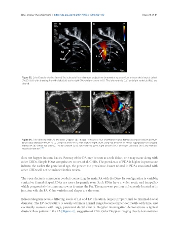
Figure 36. Two-dimensional (A) and color Doppler (B) images from apical four-chambered views demonstrating an ostium primum
atrial septal defect (Primum ASD) (long red arrow in A) with a left-to-right shunt (long red arrow in B). Mitral regurgitation (MR) jet is
marked in (B) (thick red arrow). The left atrium (LA), left ventricle (LV), right atrium (RA), and right ventricle (RV) are marked.
Modified from Ref. [24] .
does not happen in some babies. Patency of the DA may be seen as a sole defect, or it may occur along with
other CHDs. Simple PDAs comprise 6% to 11% of all CHDs. The prevalence of PDA is higher in premature
infants; the earlier the gestational age, the greater the prevalence. Issues related to PDAs associated with
other CHDs will not be included in this review.
The open ductus is a muscular conduit connecting the main PA with the DAo. Its configuration is variable;
conical or funnel shaped PDAs are more frequently seen. Such PDAs have a wider aortic end (ampulla)
which progressively becomes narrow as it enters the PA. The narrowest portion is frequently located at its
junction with the PA. Other varieties and shapes are also seen.
Echocardiogram reveals differing levels of LA and LV dilatation, largely proportional to minimal ductal
diameter. The LV contractility is usually within in normal range; becomes hyper-contractile with time, and
eventually worsens with large, persistent ductal shunts. Doppler interrogation demonstrates a typical
diastolic flow pattern in the PA [Figure 47], suggestive of PDA. Color Doppler imaging clearly demonstrates