
Page 107 - Read Online
P. 107
Rao. Vessel Plus 2022;6:25 https://dx.doi.org/10.20517/2574-1209.2021.92 Page 17 of 41
Figure 27. Transesophageal echocardiographic studies of the atrial septum demonstrating aortic (ARim), mitral (MRim), superior vena
caval (SVC Rim), and inferior vena caval (IVC Rim) rims in sort axis (A) four-chamber (B) and bi-caval (C, D) views, respectively. Aorta
(Ao), atrial septal defect (ASD), inferior vena cava (IVC), left atrium (LA), left ventricle (LV), right atrium (RA), right ventricle (RV),
and superior vena cava (SVC) are labeled.
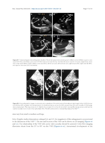
Figure 28. Echocardiographic images in subcostal echo projections of the atrial septum illustrating an atrial septal defect (ASD) (arrow
in A) before ASD occlusion (A) and placement of a buttoned device across the ASD several hours (B) and 3 months (C) following
device deployment. The long arrows in (C) identify the occluder (O) on the left atrial (LA) side and the shorter arrows point out the
counter-occluder (Co) on-end in the right atrium (RA). The left ventricle (LV) is marked. Reproduced from Ref. [27] .
sizes vary from small to medium and large.
Echo-Doppler studies demonstrate enlarged LA and LV; the magnitude of this enlargement is proportional
[33]
to the dimension of the VSD . The size and location of the VSD can be shown on 2D imaging [Figures 40
and 41]. The relationship of the VSD with aortic valve annulus should be assessed. Color flow imaging
illustrates shunt from the LV to RV via the VSD [Figures 40-44]. Aneurismal development of the