
Page 103 - Read Online
P. 103
Rao. Vessel Plus 2022;6:25 https://dx.doi.org/10.20517/2574-1209.2021.92 Page 13 of 41
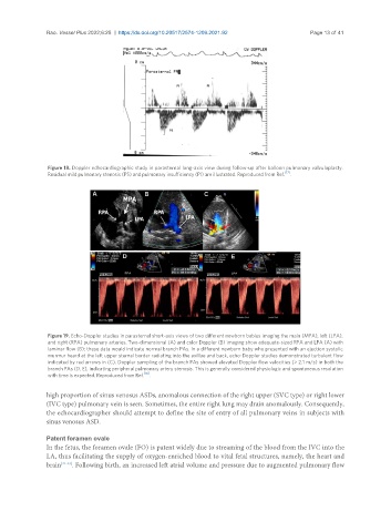
Figure 18. Doppler echocardiographic study in parasternal long-axis view during follow-up after balloon pulmonary valvuloplasty.
[17]
Residual mild pulmonary stenosis (PS) and pulmonary insufficiency (PI) are illustrated. Reproduced from Ref. .
Figure 19. Echo-Doppler studies in parasternal short-axis views of two different newborn babies imaging the main (MPA), left (LPA),
and right (RPA) pulmonary arteries. Two-dimensional (A) and color Doppler (B) imaging show adequate-sized RPA and LPA (A) with
laminar flow (B); these data would indicate normal branch PAs. In a different newborn baby who presented with an ejection systolic
murmur heard at the left upper sternal border radiating into the axillae and back, echo-Doppler studies demonstrated turbulent flow
indicated by red arrows in (C). Doppler sampling of the branch PAs showed elevated Doppler flow velocities (> 2.1 m/s) in both the
branch PAs (D, E), indicating peripheral pulmonary artery stenosis. This is generally considered physiologic and spontaneous resolution
with time is expected. Reproduced from Ref. [16] .
high proportion of sinus venosus ASDs, anomalous connection of the right upper (SVC type) or right lower
(IVC type) pulmonary vein is seen. Sometimes, the entire right lung may drain anomalously. Consequently,
the echocardiographer should attempt to define the site of entry of all pulmonary veins in subjects with
sinus venosus ASD.
Patent foramen ovale
In the fetus, the foramen ovale (FO) is patent widely due to streaming of the blood from the IVC into the
LA, thus facilitating the supply of oxygen-enriched blood to vital fetal structures, namely, the heart and
brain [21-23] . Following birth, an increased left atrial volume and pressure due to augmented pulmonary flow