
Page 105 - Read Online
P. 105
Rao. Vessel Plus 2022;6:25 https://dx.doi.org/10.20517/2574-1209.2021.92 Page 15 of 41
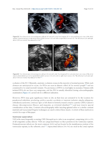
Figure 23. Two-dimensional echocardiograms without (A) and with color flow imaging (B) in subcostal long-axis view of the atrial
septum, demonstrating an atrial septal defect (ASD) (arrow in A) with left-to-right shunt (arrow in B). The left atrium (LA) and right
atrium (RA) are marked. Reproduced from Ref. [17] .
Figure 24. Two-dimensional echocardiograms without (A) and with color flow imaging (B) in subcostal short-axis views of the atrial
septum, demonstrating an atrial septal defect (ASD) (arrow in A) with a left-to-right shunt (arrow in B). The left atrium (LA) and right
atrium (RA) are marked. Reproduced from Ref. [16] .
occlusion of the FO. Ultimately, anatomic occlusion ensues in the majority of normal persons. While such
closures are anticipated to occur, the PFOs are seen in almost a third of the normal people and are
[32]
considered to be usual (normal) variants. The prevalence of PFOs is even higher in neonates. Patients with
isolated PFOs do not have any symptoms, and the PFO is usually identified during echocardiographic
examination [Figure 39] carried out for a different indication.
However, PFOs may gain significance later in life, in that they are assumed to be the location for
paradoxical embolism, producing cerebro-vascular accidents or transient ischemic attacks; platypnea-
orthodeoxia syndrome; continual right-to-left shunt in formerly treated complex cyanotic CHDs; Caisson’s
disease (decompression illness); and migraine, as reviewed elsewhere [25,31] , and may deserve special
consideration at that time. Contrast echocardiography while injecting agitated saline or a similar contrast
material and transcranial Doppler techniques are used to detect these shunts. A review of these methods is
outside the scope of this paper.
Ventricular septal defect
VSD is the most frequently occurring CHD (bicuspid aortic valve is an exception), comprising 20% to 25%
of all congenital cardiac defects. VSDs are categorized based on their position in the ventricular septum.
Perimembranous defects are the most common (80%) and are located in the membranous portion of the
ventricular septum, in the subaortic area [3,24] . Supracristal defects (5%-7%) are sited in the conal septum