
Page 110 - Read Online
P. 110
Page 20 of 41 Rao. Vessel Plus 2022;6:25 https://dx.doi.org/10.20517/2574-1209.2021.92
Figure 33. Echo-Doppler studies in subcostal projections of the atrial septum with color flow Doppler illustrating the buttoned device
(unfilled arrowheads) across atrial septal defects in two children (A) and (B) with residual shunts (filled arrowheads). The left atrium
(LA) and right atrium (RA) are marked. Reproduced from Ref. [31] .
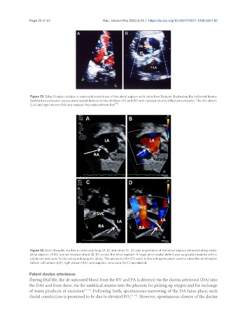
Figure 34. Echo-Doppler studies in subcostal long (A, B) and short (C, D) axis projections of the atrial septum demonstrating intact
atrial septum (A-D) and no residual shunt (B, D) across the atrial septum. A large atrial septal defect was surgically repaired with a
patch one year prior to this echocardiographic study. The arrows in (A)-(D) point to the echogenic patch used to close the atrial septal
defect. Left atrium (LA), right atrium (RA), and superior vena cava (SVC) are labeled.
Patent ductus arteriosus
During fetal life, the de-saturated blood from the RV and PA is diverted via the ductus arteriosus (DA) into
the DAo and from there, via the umbilical arteries into the placenta for picking up oxygen and for exchange
of waste products of excretion [21-23] . Following birth, spontaneous narrowing of the DA takes place; such
ductal constriction is presumed to be due to elevated PO 2 [21-23] . However, spontaneous closure of the ductus