
Page 102 - Read Online
P. 102
Page 12 of 41 Rao. Vessel Plus 2022;6:25 https://dx.doi.org/10.20517/2574-1209.2021.92
Figure 16. Doppler echocardiographic studies in parasternal long-axis view before (A), the day after (B), and 8 months (C) following
balloon pulmonary valvuloplasty (BPV), illustrating a marked reduction in the peak Doppler flow velocity and peak instantaneous
gradient from 92 mmHg to 17 mmHg immediately following BPV (A, B). The residual gradient was small (20 mmHg) eight months after
BPV (C). Reproduced from Ref. [18] .
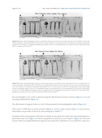
Figure 17. Doppler echocardiographic studies in parasternal long-axis view before (A), the day after (B), and 10 months after (C)
balloon pulmonary valvuloplasty (BPV). While there was a substantial reduction in peak Doppler flow velocity (and calculated gradients
from 120 mmHg to 48 mmHg) immediately following BPV (A vs. B), there was a residual 48 mmHg gradient; more importantly
distinctive triangular pattern, indicative of infundibular obstruction remained (B). The residual gradient at ten months following BPV
was low (25 mmHg) without the triangular pattern, suggesting that the infundibular narrowing has resolved (C). The residual peak
[18]
gradients are printed at the lower portion of each panel. Reproduced from Ref. .
Echocardiography is also useful in demonstrating the effectiveness of device occlusion [Figures 28-32] and
detecting residual shunts [Figure 33].
The effectiveness of surgical closure can also be demonstrated in echocardiographic studies [Figure 34].
Other types of ASDs such as ostium primum [Figures 35 and 36], sinus venosus [Figure 37], and coronary
sinus defects can generally be shown by echocardiographic studies [23,24] .
In patients with ostium primum ASD, there is usually an associated cleft mitral valve, best demonstrated in
apical short-axis view [Figure 38]. Mitral regurgitation is shown by color Doppler [Figure 36]. The sinus
venosus ASDs are usually divided into superior vena caval (SVC) and inferior vena caval (IVC) types. In a