
Page 96 - Read Online
P. 96
Page 6 of 41 Rao. Vessel Plus 2022;6:25 https://dx.doi.org/10.20517/2574-1209.2021.92
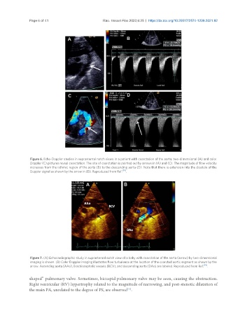
Figure 6. Echo-Doppler studies in suprasternal notch views in a patient with coarctation of the aorta; two-dimensional (A) and color
Doppler (C) pictures reveal coarctation. The site of coarctation is pointed out by arrows in (A) and (C). The magnitude of flow velocity
increases from the isthmic region of the aorta (B) to the descending aorta (D). Note that there is extension into the diastole of the
Doppler signal as shown by the arrow in (D). Reproduced from Ref. [10] .
Figure 7. (A) Echocardiographic study in suprasternal notch view of a baby with coarctation of the aorta (arrow) by two-dimensional
imaging is shown. (B) Color Doppler imaging illustrates flow turbulence at the location of the coarcted aortic segment as shown by the
arrow. Ascending aorta (AAo), Brachiocephalic vessels (BCV), and descending aorta (DAo) are labeled. Reproduced from Ref. [10] .
shaped” pulmonary valve. Sometimes, bicuspid pulmonary valve may be seen, causing the obstruction.
Right ventricular (RV) hypertrophy related to the magnitude of narrowing, and post-stenotic dilatation of
the main PA, unrelated to the degree of PS, are observed .
[15]