
Page 92 - Read Online
P. 92
Page 2 of 41 Rao. Vessel Plus 2022;6:25 https://dx.doi.org/10.20517/2574-1209.2021.92
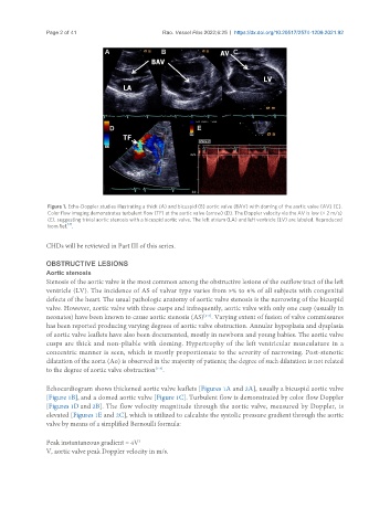
Figure 1. Echo-Doppler studies illustrating a thick (A) and bicuspid (B) aortic valve (BAV) with doming of the aortic valve (AV) (C).
Color flow imaging demonstrates turbulent flow (TF) at the aortic valve (arrow) (D). The Doppler velocity via the AV is low (< 2 m/s)
(E), suggesting trivial aortic stenosis with a bicuspid aortic valve. The left atrium (LA) and left ventricle (LV) are labeled. Reproduced
[4]
from Ref. .
CHDs will be reviewed in Part III of this series.
OBSTRUCTIVE LESIONS
Aortic stenosis
Stenosis of the aortic valve is the most common among the obstructive lesions of the outflow tract of the left
ventricle (LV). The incidence of AS of valvar type varies from 5% to 6% of all subjects with congenital
defects of the heart. The usual pathologic anatomy of aortic valve stenosis is the narrowing of the bicuspid
valve. However, aortic valve with three cusps and infrequently, aortic valve with only one cusp (usually in
[1-3]
neonates) have been known to cause aortic stenosis (AS) . Varying extent of fusion of valve commissures
has been reported producing varying degrees of aortic valve obstruction. Annular hypoplasia and dysplasia
of aortic valve leaflets have also been documented, mostly in newborn and young babies. The aortic valve
cusps are thick and non-pliable with doming. Hypertrophy of the left ventricular musculature in a
concentric manner is seen, which is mostly proportionate to the severity of narrowing. Post-stenotic
dilatation of the aorta (Ao) is observed in the majority of patients; the degree of such dilatation is not related
to the degree of aortic valve obstruction .
[1-3]
Echocardiogram shows thickened aortic valve leaflets [Figures 1A and 2A], usually a bicuspid aortic valve
[Figure 1B], and a domed aortic valve [Figure 1C]. Turbulent flow is demonstrated by color flow Doppler
[Figures 1D and 2B]. The flow velocity magnitude through the aortic valve, measured by Doppler, is
elevated [Figures 1E and 2C], which is utilized to calculate the systolic pressure gradient through the aortic
valve by means of a simplified Bernoulli formula:
Peak instantaneous gradient = 4V
2
V, aortic valve peak Doppler velocity in m/s.