
Page 87 - Read Online
P. 87
Page 20 of 23 Rao. Vessel Plus 2022;6:24 https://dx.doi.org/10.20517/2574-1209.2021.91
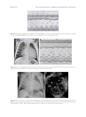
Figure 27. M-mode echocardiogram of a baby who had cardiomegaly on a chest X-ray demonstrated a severely enlarged left ventricle
(LV) with markedly diminished LV systolic function (shortening fraction by m-mode echo was 13%).
Figure 28. Chest roentgenogram (A) exhibits cardiac enlargement. M-mode echo cardiogram (B) of the same baby demonstrates
dilatation of the left ventricle (LV), thought to be secondary to dilated cardiomyopathy.
Figure 29. Chest X-ray (A) of a neonate demonstrating severe cardiomegaly who on a two-dimensional echocardiogram (B) revealed
Ebstein’s anomaly of the tricuspid valve [arrow in (B)] with marked tricuspid insufficiency (not shown). Arrows in the (A) demarcate
enlarged right atrium (RA). aRV: Atrialized right ventricle; LA: left atrium; LV: left ventricle; RV: right ventricle.