
Page 86 - Read Online
P. 86
Rao. Vessel Plus 2022;6:24 https://dx.doi.org/10.20517/2574-1209.2021.91 Page 19 of 23
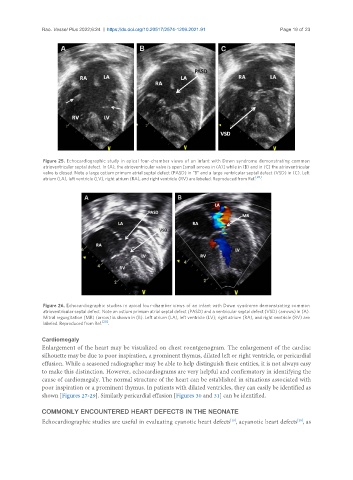
Figure 25. Echocardiographic study in apical four-chamber views of an infant with Down syndrome demonstrating common
atrioventricular septal defect. In (A), the atrioventricular valve is open [small arrows in (A)] while in (B) and in (C) the atrioventricular
valve is closed. Note a large ostium primum atrial septal defect (PASD) in “B” and a large ventricular septal defect (VSD) in (C). Left
[20]
atrium (LA), left ventricle (LV), right atrium (RA), and right ventricle (RV) are labeled. Reproduced from Ref. .
Figure 26. Echocardiographic studies in apical four-chamber views of an infant with Down syndrome demonstrating common
atrioventricular septal defect. Note an ostium primum atrial septal defect (PASD) and a ventricular septal defect (VSD) (arrows) in (A).
Mitral regurgitation (MR) (arrow) is shown in (B). Left atrium (LA), left ventricle (LV); right atrium (RA), and right ventricle (RV) are
labeled. Reproduced from Ref. [20] .
Cardiomegaly
Enlargement of the heart may be visualized on chest roentgenogram. The enlargement of the cardiac
silhouette may be due to poor inspiration, a prominent thymus, dilated left or right ventricle, or pericardial
effusion. While a seasoned radiographer may be able to help distinguish these entities, it is not always easy
to make this distinction. However, echocardiograms are very helpful and confirmatory in identifying the
cause of cardiomegaly. The normal structure of the heart can be established in situations associated with
poor inspiration or a prominent thymus. In patients with dilated ventricles, they can easily be identified as
shown [Figures 27-29]. Similarly pericardial effusion [Figures 30 and 31] can be identified.
COMMONLY ENCOUNTERED HEART DEFECTS IN THE NEONATE
Echocardiographic studies are useful in evaluating cyanotic heart defects , acyanotic heart defects , as
[20]
[19]