
Page 81 - Read Online
P. 81
Page 14 of 23 Rao. Vessel Plus 2022;6:24 https://dx.doi.org/10.20517/2574-1209.2021.91
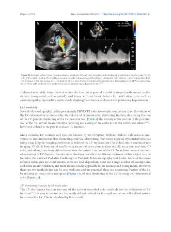
Figure 19. Selected video frames from parasternal short-axis 2D and color Doppler study displaying a patent ductus arteriosus (PDA)
with left-to-right shunt in (A). Continuous wave Doppler interrogation of the PDA in (B) depicts high velocity (~4 m/s) indicating that
the pressure in the pulmonary artery is likely to be low (see the text). Aorta (Ao), diastolic (D), descending aorta (DAo), pulmonary
[20]
artery (PA), right ventricle (RV), and systolic (S) are labeled. Reproduced from Ref. .
addressed separately. Assessment of ventricular function is generally useful in subjects with known cardiac
defects (congenital and acquired) and those without heart defects but with situations such as
cardiomyopathy, myocarditis, septic shock, diaphragmatic hernia, and persistent pulmonary hypertension.
Left ventricle
Several echocardiographic techniques, namely PEP/LVET ratio, isovolumic contraction time, the volume of
the LV calculated by m-mode echo, the velocity of circumferential shortening fraction, shortening fraction
of the LV, percent thickening of the LV posterior wall [Table 4], the velocity of the motion of the posterior
wall of the LV, several measurements of opening and closing of the aortic and mitral valves, and others [22,23] ,
have been utilized in the past to evaluate LV function.
More recently, LV volumes and ejection fraction by 2D (Simpson, Biplane, Bullet), wall stress at end-
systole-to-circumferential fiber shortening, mid-wall shortening, fiber stress, regional myocardial velocities
using tissue Doppler imaging, performance index of the LV myocardium (Tei index), strain and strain rate
imaging, LV dP/dt from mitral insufficiency jet, mitral valve annular plane systolic excursion, real-time 3D
echo, and others, have been utilized to evaluate the systolic function of the LV. In addition, several methods
of evaluation of LV diastolic function have also been described. Additional treatment of this subject may be
found in the standard Pediatric Cardiology or Pediatric Echocardiography text books. Some of the above
referred techniques are cumbersome, some are load-dependent, some use a large number of assumptions,
and some are not validated, and most are not readily applicable to the neonate and young infant. However,
there are two methods that can be used with ease and are practical; these are shortening fraction of the LV
by utilizing m-mode echocardiogram [Figure 1] and area shortening of the LV by using two-dimensional
echo [Figure 20].
LV shortening fraction by M-mode echo
The LV shortening fraction was one of the earliest described echo methods for the evaluation of LV
[24]
function . It is easy to use and is a frequently utilized method for the rapid evaluation of the global systolic
function of the LV. This is calculated by the formula: