
Page 77 - Read Online
P. 77
Page 10 of 23 Rao. Vessel Plus 2022;6:24 https://dx.doi.org/10.20517/2574-1209.2021.91
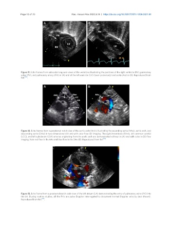
Figure 11. Echo frames from subcostal long-axis views of the ventricles illustrating the positions of the right ventricle (RV), pulmonary
valve (PV), and pulmonary artery (PA) in (A) and of the left ventricle (LV) (seen posteriorly) and aorta (Ao) in (B). Reproduced from
Ref. [19] .
Figure 12. Echo frames from suprasternal notch view of the aortic arch (Arch) illustrating the ascending aorta (AAo), aortic arch, and
descending aorta (DAo) in two-dimensional (A) and with color flow (B) imaging. The right innominate (RInn), left common carotid
(LCC), and left subclavian (LSA) arteries originating from the aortic arch are demonstrated without in (A) and with color in (B) flow
imaging. Note red flow in the AAo and blue flow in the DAo (B). Reproduced from Ref. [19] .
Figure 13. Echo frame from suprasternal notch crab-view of the left atrium (LA) demonstrating the entry of pulmonary veins (PV) into
the LA. During routine studies, all the PVs are pulse Doppler interrogated to document normal Doppler velocity (not shown).
Reproduced from Ref. [19] .