
Page 74 - Read Online
P. 74
Rao. Vessel Plus 2022;6:24 https://dx.doi.org/10.20517/2574-1209.2021.91 Page 7 of 23
Table 2. Normal intra-cardiac velocities by Doppler ultrasound*
Site Children m/s; mean (range) Adults m/s; mean (range)
Mitral inflow 1.00 (0.8-1.3) 0.90 (0.6-1.3)
Tricuspid inflow 0.60 (0.5-0.8) 0.50 (0.3-0.7)
Pulmonary artery 0.90 (0.7-1.1) 0.75 (0.6-0.9)
Left ventricle 1.00 (0.7-1.2) 0.90 (0.7-1.1)
Aorta 1.50 (1.2-1.8) 1.35 (1.0-1.7)
*Data abstracted from Refs. [15,16] .
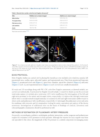
Figure 7. Two-dimensional echo and color Doppler video frames are presented to demonstrate turbulent flow patterns. (A) Turbulent
blood flow (arrow) commences at the level of the aortic valve (arrow - TF) because of aortic stenosis while (B) the turbulence begins at
the ductus (TF) shown by arrow. Aorta (Ao), descending aorta (DAo), left ventricle (LV), and main pulmonary artery (MPA) are
labeled. Reproduced from Ref. [19] .
ECHO PROTOCOL
Echo-Doppler studies are carried out by placing the transducer over multiple echo windows, namely, left
parasternal area, cardiac apex, subcostal region, and suprasternal area. Data from parasternal long-axis
[Figure 2], parasternal short-axis [Figure 8], apical four-chamber, apical five-chamber [Figure 9], subcostal
[Figures 10 and 11], and suprasternal notch [Figures 12 and 13] views are usually documented.
M-mode and 2D recordings along with PW, CW, color flow Doppler assessment, as deemed suitable, are
carried out methodically. Examination by Doppler should include (1) search for shunts across the atrial and
ventricular septae; (2) exclude atrio-ventricular (AV) valve insufficiency by interrogation of the left and
right atria proximal to the mitral and tricuspid valves, respectively; (3) explore the left/right ventricular
inflow sites to exclude mitral/tricuspid stenosis; (4) examine the left and right ventricular outflow tracts to
detect aortic and pulmonary valve insufficiency, respectively; (5) interrogate the pulmonary artery and aorta
for semilunar valve stenosis; and (6) examination looking for aortic coarctation and patency of the ductus
arteriosus. Investigation of additional sites on the basis of findings of clinical, laboratory and
echocardiographic examination should be performed as well.
METHODS OF ESTIMATION OF PULMONARY ARTERY PRESSURE
Frequently, neonatologists, pediatric cardiologists, pediatric intensivists, cardiac surgeons and pediatricians
request for evaluation of PA pressures in their patients. Although the reasons for such request vary from
one specialist to the other, the magnitude of the PA pressure is a central component in managing many