
Page 75 - Read Online
P. 75
Page 8 of 23 Rao. Vessel Plus 2022;6:24 https://dx.doi.org/10.20517/2574-1209.2021.91
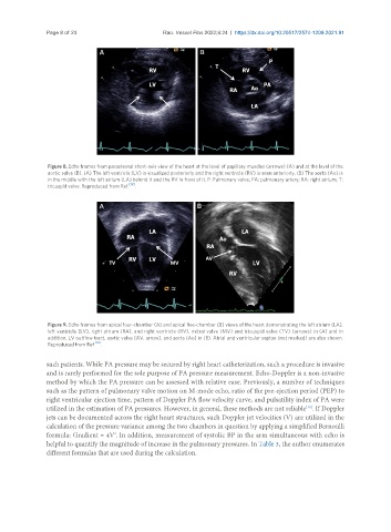
Figure 8. Echo frames from parasternal short-axis view of the heart at the level of papillary muscles (arrows) (A) and at the level of the
aortic valve (B). (A) The left ventricle (LV) is visualized posteriorly and the right ventricle (RV) is seen anteriorly. (B) The aorta (Ao) is
in the middle with the left atrium (LA) behind it and the RV in front of it. P: Pulmonary valve; PA: pulmonary artery; RA: right atrium; T:
[19]
tricuspid valve. Reproduced from Ref. .
Figure 9. Echo frames from apical four-chamber (A) and apical five-chamber (B) views of the heart demonstrating the left atrium (LA),
left ventricle (LV), right atrium (RA), and right ventricle (RV), mitral valve (MV) and tricuspid valve (TV) (arrows) in (A) and in
addition, LV outflow tract, aortic valve (AV, arrow), and aorta (Ao) in (B). Atrial and ventricular septae (not marked) are also shown.
Reproduced from Ref. [19] .
such patients. While PA pressure may be secured by right heart catheterization, such a procedure is invasive
and is rarely performed for the sole purpose of PA pressure measurement. Echo-Doppler is a non-invasive
method by which the PA pressure can be assessed with relative ease. Previously, a number of techniques
such as the pattern of pulmonary valve motion on M-mode echo, ratio of the pre-ejection period (PEP) to
right ventricular ejection time, pattern of Doppler PA flow velocity curve, and pulsatility index of PA were
utilized in the estimation of PA pressures. However, in general, these methods are not reliable . If Doppler
[19]
jets can be documented across the right heart structures, such Doppler jet velocities (V) are utilized in the
calculation of the pressure variance among the two chambers in question by applying a simplified Bernoulli
formula: Gradient = 4V . In addition, measurement of systolic BP in the arm simultaneous with echo is
2
helpful to quantify the magnitude of increase in the pulmonary pressures. In Table 3, the author enumerates
different formulas that are used during the calculation.