
Page 84 - Read Online
P. 84
Rao. Vessel Plus 2022;6:24 https://dx.doi.org/10.20517/2574-1209.2021.91 Page 17 of 23
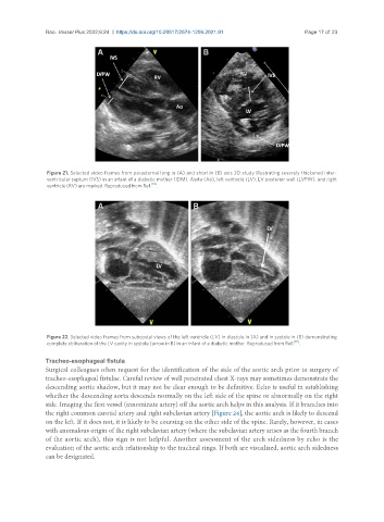
Figure 21. Selected video frames from parasternal long in (A) and short in (B) axis 2D study illustrating severely thickened inter-
ventricular septum (IVS) in an infant of a diabetic mother (IDM). Aorta (Ao), left ventricle (LV), LV posterior wall (LVPW), and right
ventricle (RV) are marked. Reproduced from Ref. [19] .
Figure 22. Selected video frames from subcostal views of the left ventricle (LV) in diastole in (A) and in systole in (B) demonstrating
complete obliteration of the LV cavity in systole (arrow in B) in an infant of a diabetic mother. Reproduced from Ref. [19] .
Tracheo-esophageal fistula
Surgical colleagues often request for the identification of the side of the aortic arch prior to surgery of
tracheo-esophageal fistulae. Careful review of well penetrated chest X-rays may sometimes demonstrate the
descending aortic shadow, but it may not be clear enough to be definitive. Echo is useful in establishing
whether the descending aorta descends normally on the left side of the spine or abnormally on the right
side. Imaging the first vessel (innominate artery) off the aortic arch helps in this analysis. If it branches into
the right common carotid artery and right subclavian artery [Figure 24], the aortic arch is likely to descend
on the left. If it does not, it is likely to be coursing on the other side of the spine. Rarely, however, in cases
with anomalous origin of the right subclavian artery (where the subclavian artery arises as the fourth branch
of the aortic arch), this sign is not helpful. Another assessment of the arch sidedness by echo is the
evaluation of the aortic arch relationship to the tracheal rings. If both are visualized, aortic arch sidedness
can be designated.