
Page 88 - Read Online
P. 88
Rao. Vessel Plus 2022;6:24 https://dx.doi.org/10.20517/2574-1209.2021.91 Page 21 of 23
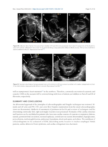
Figure 30. Selected video frames from apical four-chamber [(A) and (B)] and parasternal short-axis (C) projections of the heart in a
baby with cardiac enlargement on a chest X-ray demonstrating a large pericardial effusion (thick arrows). Thin arrows mark fibrin
strands within the effusion. Reproduced from Ref. [19] .
Figure 31. Selected video frames from parasternal long in (A) and short in (B) axis views in an infant with cardiac enlargement on chest
X-ray demonstrate a large pericardial effusion (arrows). Reproduced from Ref. [19] .
well as asymptomatic heart murmurs in the newborn. Therefore, commonly encountered acyanotic and
[34]
cyanotic CHDs in the neonate will be reviewed along with those of infants and children in Parts II and III of
this series, respectively.
SUMMARY AND CONCLUSIONS
An abbreviated appraisal of the principles of echocardiographic and Doppler techniques was reviewed. M-
mode and 2D echo and PW, CW, and color flow Doppler examination from the usual echocardiographic
views are documented. Methods of assessment of pressures in the PA and a review of techniques used for
determining ventricular (left, right and single) function were presented. Normality of the cardiac structure
and function can be established in patients who have non-cardiac reasons of cyanosis or respiratory distress,
namely, persistent fetal circulation, neonatal asphyxia, central nervous system abnormalities, hypoglycemia,
polycythemia, methemoglobinemia, pulmonary hypoplasia, shock and sepsis, and others. The usefulness of
echocardiograms in the evaluation of IDM, descending aortic location in tracheo-esophageal fistula
patients, cardiac defects in Down syndrome, and cardiac enlargement was elucidated.