
Page 47 - Read Online
P. 47
Uppu. Vessel Plus 2021;6:21 https://dx.doi.org/10.20517/2574-1209.2021.101 Page 11 of 19
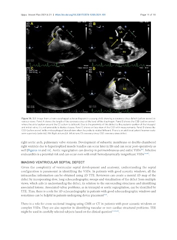
Figure 14. Still image from a trans-esophageal echocardiogram in a young child showing a coronary sinus defect (yellow arrow) in
various views. Panel A shows the length of the coronary sinus at the level of the diaphragm. Panel B shows the CSD (yellow arrow)
where the atrial septum around the CS ostium is deficient. Due to the proximity of this defect to the posterior portion of the tricuspid
and mitral valve, it is not amenable to device closure. Panel C shows en face view of the CSD with measurements. Panel D shows the
CSD (yellow arrow) in the midesophageal bicaval view when the probe is rotated leftward. There is an additional patent foramen ovale
seen superiorly (asterisk). RA: Right atrium; LA: left atrium; CS: coronary sinus; CSD: coronary sinus defect.
right aortic arch, pulmonary valve stenosis. Development of subaortic membrane or double-chambered
right ventricle due to hypertrophied muscle bundles can occur later in life and can recur post-operatively as
well [Figures 18 and 19]. Aortic regurgitation can develop in perimembranous and outlet VSDs . Infective
[41]
endocarditis is a potential risk and can occur even with small hemodynamically insignificant VSDs [10,42] .
IMAGING VENTRICULAR SEPTAL DEFECT
Given the complexity of ventricular septal development and anatomy, understanding the septal
configuration is paramount in identifying the VSDs. In patients with good acoustic windows, all the
intracardiac information can be obtained using 2D TTE. Reviewers can create a mental 3D map of the
defect by incorporating slow, long echocardiographic sweeps and visualization of the defect from multiple
views, which aids in understanding the defect, its relation to the surrounding structures and identifying
associated lesions. Associated valvar problems, as in tricuspid or aortic regurgitation, can be identified by
TTE. Thus, there is a role for 3D echocardiography in patients with good echocardiographic windows and
sometimes can be helpful in patients undergoing device placement .
[5,6]
There is a role for cross-sectional imaging using CMR or CT in patients with poor acoustic windows or
complex VSDs. They are also superior in identifying vascular or non-cardiac structural problems. TEE
might be used in carefully selected subjects based on the clinical question [13,23,43] .