
Page 50 - Read Online
P. 50
Page 14 of 19 Uppu. Vessel Plus 2021;6:21 https://dx.doi.org/10.20517/2574-1209.2021.101
Figure 19. Still trans-esophageal echocardiogram in a young child in long-axis view showing a discrete membrane (red arrow) within the
left ventricular outflow tract below the aortic valve. LA: Left atrium; LV: left ventricle.
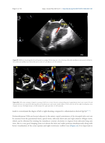
Figure 20. Still color compare image in a young child from a trans-thoracic echocardiogram in parasternal short axis (panel A) and
Subcostal short axis (panel B) showing the perimembranous ventricular septal defect (VSD) with the left to right shunting by color
Doppler (arrow). RA: Right atrium; LA: left atrium; RV: right ventricle; LV: left ventricle.
tends to overestimate the degree of left to right shunting compared to catheterization derived Qp/Qs [9,44-46] .
Perimembranous VSDs are located adjacent to the antero-septal commissure of the tricuspid valve and can
be assessed from the parasternal views, apical views, subcostal short axis and right anterior oblique views,
which can be obtained by rotating the transducer counter-clockwise 45 degrees from subcostal long-axis
view, this is a very good imaging plane as it shows the inlet and outlet portions simultaneously along with
better visualization of the conal septum and right ventricular outflow tract [Figure 20]. It is important to