
Page 48 - Read Online
P. 48
Page 12 of 19 Uppu. Vessel Plus 2021;6:21 https://dx.doi.org/10.20517/2574-1209.2021.101
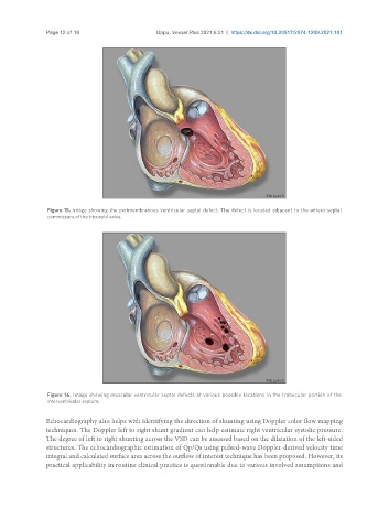
Figure 15. Image showing the perimembranous ventricular septal defect. The defect is located adjacent to the antero-septal
commissure of the tricuspid valve.
Figure 16. Image showing muscular ventricular septal defects at various possible locations in the trabecular portion of the
interventricular septum.
Echocardiography also helps with identifying the direction of shunting using Doppler color flow mapping
techniques. The Doppler left to right shunt gradient can help estimate right ventricular systolic pressure.
The degree of left to right shunting across the VSD can be assessed based on the dilatation of the left-sided
structures. The echocardiographic estimation of Qp/Qs using pulsed-wave Doppler-derived velocity time
integral and calculated surface area across the outflow of interest technique has been proposed. However, its
practical applicability in routine clinical practice is questionable due to various involved assumptions and