
Page 51 - Read Online
P. 51
Uppu. Vessel Plus 2021;6:21 https://dx.doi.org/10.20517/2574-1209.2021.101 Page 15 of 19
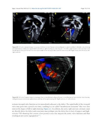
Figure 21. Still color compare image in a young child from a trans-thoracic echocardiogram in apical modified 4-chamber view showing
perimembranous ventricular septal defect with left to right shunting by color Doppler. The left to right shunt is restricted due to the
membranous aneurysm formed from the septal leaflet of the tricuspid valve (asterisk and arrow). RA: Right atrium; LA: left atrium; RV:
right ventricle.
Figure 22. Still color Doppler image in a teenager from a trans-thoracic echocardiogram in modified apical four-chamber view showing
multiple muscular ventricular septal defects with the left to right shunting. RV: Right ventricle; LV: left ventricle.
evaluate tricuspid valve function as it is immediately adjacent to the defect. The septal leaflet of the tricuspid
valve may grow into a pouch over time, resulting in a so-called “membranous aneurysm” that over time
restricts the degree of left to right shunting [Figure 21]. In addition, the aortic right and non-coronary cusps
are directly above the defect and sometimes may prolapse into the defect due to lack of support and high-
velocity VSD shunting that creates a low-pressure zone that impacts the aortic valve function and thus
resulting in new aortic regurgitation [3,41] .