
Page 49 - Read Online
P. 49
Uppu. Vessel Plus 2021;6:21 https://dx.doi.org/10.20517/2574-1209.2021.101 Page 13 of 19
Figure 17. Image showing outlet ventricular septal defect. The defect is located below the pulmonary valve.
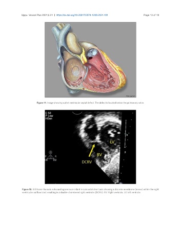
Figure 18. Still trans-thoracic echocardiogram in an infant in subcostal short axis showing a discrete membrane (arrow) within the right
ventricular outflow tract resulting in a double-chambered right ventricle (DCRV). RV: Right ventricle; LV: left ventricle.