
Page 45 - Read Online
P. 45
Uppu. Vessel Plus 2021;6:21 https://dx.doi.org/10.20517/2574-1209.2021.101 Page 9 of 19
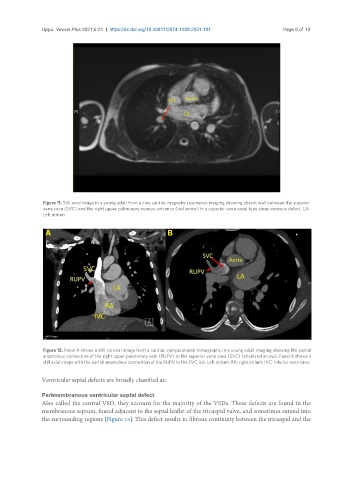
Figure 11. Still axial image in a young adult from a cine cardiac magnetic resonance imaging showing absent wall between the superior
vena cava (SVC) and the right upper pulmonary venous entrance (red arrow) in a superior vena caval type sinus venosus defect. LA:
Left atrium.
Figure 12. Panel A shows a still coronal image from a cardiac computational tomography in a young adult imaging showing the partial
anomalous connection of the right upper pulmonary vein (RUPV) to the superior vena cava (SVC) (short red arrow). Panel B shows a
still axial image with the partial anomalous connection of the RUPV to the SVC. LA: Left atrium; RA: right atrium; IVC: inferior vena cava.
Ventricular septal defects are broadly classified as:
Perimembranous ventricular septal defect
Also called the central VSD, they account for the majority of the VSDs. These defects are found in the
membranous septum, found adjacent to the septal leaflet of the tricuspid valve, and sometimes extend into
the surrounding regions [Figure 15]. This defect results in fibrous continuity between the tricuspid and the