
Page 40 - Read Online
P. 40
Page 4 of 19 Uppu. Vessel Plus 2021;6:21 https://dx.doi.org/10.20517/2574-1209.2021.101
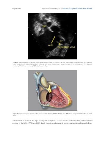
Figure 3. Still image from a trans-thoracic echocardiogram in subcostal short-axis view in a teenager showing a large IVC confluent
ostium secundum atrial septal defect (long yellow arrow), notice the prominent Eustachian valve (short yellow arrow). SVC: Superior
vena cava; IVC: inferior vena cava; ASD: atrial septal defect.
Figure 4. Image showing the location of the ostium primum atrial septal defect at the crux of the heart along with inlet ventricular septal
defect.
communication between the right-sided pulmonary veins and the cardiac end of the SVC or the superior
portion of the RA in SVC type SVD. Rarely there is a deficiency of wall separating the right middle/lower