
Page 43 - Read Online
P. 43
Uppu. Vessel Plus 2021;6:21 https://dx.doi.org/10.20517/2574-1209.2021.101 Page 7 of 19
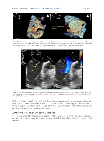
Figure 7. Still three-dimensional images from a trans-esophageal echocardiogram performed during device placement in a teenager,
panel A shows an ostium secundum atrial septal defect with deficient retroaortic rim (red arrow). Panel B shows a successfully deployed
GORE® CARDIOFORM ASD occluder in the satisfactory position seen in its entirety (red arrow). ASD: Atrial septal defect.
Figure 8. Still image from a trans-esophageal echocardiogram in a young adult showing an ostium secundum atrial septal defect with
deficient retroaortic rim (yellow arrow), color flow mapping shows left to right shunting across the defect. RA: Right atrium; LA: left
atrium; ASD: atrial septal defect.
CSDs are difficult to visualize by traditional views. It is important to perform echo sweeps to image the
atrial septum carefully around the location of the CS ostium, and TEE may help in those with difficult
windows [Figure 14]. Contrast injection in the left arm in patients with suspected CSD with partially or
completely unroofed CS can help with identification .
[7]
ANATOMY OF VENTRICULAR SEPTAL DEFECTS
The ventricular septal formation is a complex process that begins at the end of the 4th week. Therefore, a
detailed description of the ventricular septal formation, including genetic basis, is beyond the scope of this
chapter [3,5,12,34,35] .