
Page 44 - Read Online
P. 44
Page 8 of 19 Uppu. Vessel Plus 2021;6:21 https://dx.doi.org/10.20517/2574-1209.2021.101
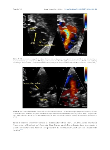
Figure 9. Still color compare image from a trans-thoracic echocardiogram in a young child in subcostal short-axis view showing a
superior vena caval type sinus venosus defect (long yellow arrow). Flow from the right upper pulmonary vein (RUPV) is seen emptying
into the right atrium. There is a prominent Eustachian valve (short yellow arrow) seen anterior to the inferior vena caval entrance (IVC).
Figure 10. Still color compare image from a trans-thoracic echocardiogram in a young child in high right parasternal short-axis view
showing an inferior vena caval type sinus venosus defect behind the prominent Eustachian valve (short yellow arrow). Flow from the
right lower pulmonary vein (Rt LPV) is seen emptying into the right atrium adjacent to the entrance of the inferior vena caval entrance
(IVC).
There is extensive controversy around the nomenclature of the VSDs. The International Society for
Nomenclature of Paediatric and Congenital Heart Disease has tried to address the issue by proposing a
classification scheme that has been incorporated in the International Classification of Diseases-11th
iteration [11,36] .