
Page 46 - Read Online
P. 46
Page 10 of 19 Uppu. Vessel Plus 2021;6:21 https://dx.doi.org/10.20517/2574-1209.2021.101
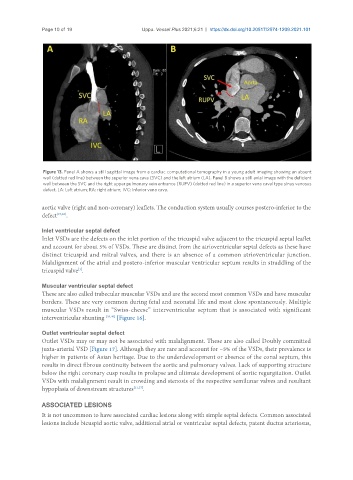
Figure 13. Panel A shows a still sagittal image from a cardiac computational tomography in a young adult imaging showing an absent
wall (dotted red line) between the superior vena cava (SVC) and the left atrium (LA). Panel B shows a still axial image with the deficient
wall between the SVC and the right upper pulmonary vein entrance (RUPV) (dotted red line) in a superior vena caval type sinus venosus
defect. LA: Left atrium; RA: right atrium; IVC: inferior vena cava.
aortic valve (right and non-coronary) leaflets. The conduction system usually courses postero-inferior to the
defect [37,38] .
Inlet ventricular septal defect
Inlet VSDs are the defects on the inlet portion of the tricuspid valve adjacent to the tricuspid septal leaflet
and account for about 5% of VSDs. These are distinct from the atrioventricular septal defects as these have
distinct tricuspid and mitral valves, and there is an absence of a common atrioventricular junction.
Malalignment of the atrial and postero-inferior muscular ventricular septum results in straddling of the
[3]
tricuspid valve .
Muscular ventricular septal defect
These are also called trabecular muscular VSDs and are the second most common VSDs and have muscular
borders. These are very common during fetal and neonatal life and most close spontaneously. Multiple
muscular VSDs result in “Swiss-cheese” interventricular septum that is associated with significant
interventricular shunting [39,40] [Figure 16].
Outlet ventricular septal defect
Outlet VSDs may or may not be associated with malalignment. These are also called Doubly committed
juxta-arterial VSD [Figure 17]. Although they are rare and account for ~5% of the VSDs, their prevalence is
higher in patients of Asian heritage. Due to the underdevelopment or absence of the conal septum, this
results in direct fibrous continuity between the aortic and pulmonary valves. Lack of supporting structure
below the right coronary cusp results in prolapse and ultimate development of aortic regurgitation. Outlet
VSDs with malalignment result in crowding and stenosis of the respective semilunar valves and resultant
hypoplasia of downstream structures [11,37] .
ASSOCIATED LESIONS
It is not uncommon to have associated cardiac lesions along with simple septal defects. Common associated
lesions include bicuspid aortic valve, additional atrial or ventricular septal defects, patent ductus arteriosus,