
Page 42 - Read Online
P. 42
Page 6 of 19 Uppu. Vessel Plus 2021;6:21 https://dx.doi.org/10.20517/2574-1209.2021.101
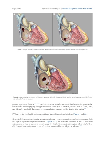
Figure 5. Images showing superior vena caval (A) and inferior vena caval type (B) of sinus venosus defects, respectively.
Figure 6. Image showing the location of the coronary sinus defect (yellow asterisk) in relation to ostium secundum ASD (green
asterisk). ASD: Atrial septal defect.
provide superior 3D datasets [10,13,23,24] . Furthermore, CMR provides additional data by quantifying ventricular
volumes and obtaining Qp/Qs using phase contrast techniques. In addition, datasets from 3D echo, CMR,
[32]
and CT can be fused with fluoroscopy to reduce radiation exposure and the time for intervention .
SVDs are better visualized from the subcostal and high right parasternal windows [Figures 9 and 10].
Given the high association of partial anomalous pulmonary venous connections, one has to consider a CMR
or CT prior to planned surgical intervention [Figures 11-13]. Transcatheter correction of the SVC type SVD
using a covered stent is feasible in a select group of patients. Cross-sectional imaging, either with CMR or
CT, along with simulation using virtual 3D models, is essential for careful patient selection [9,33] .