
Page 156 - Read Online
P. 156
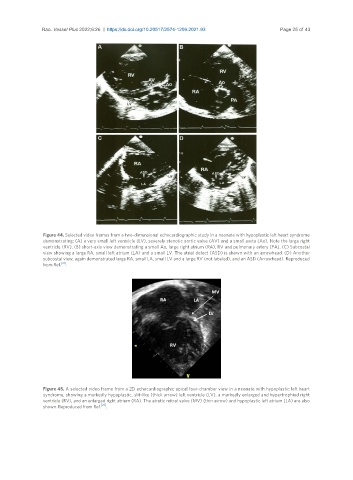
Rao. Vessel Plus 2022;6:26  https://dx.doi.org/10.20517/2574-1209.2021.93       Page 25 of 43


                Figure 44. Selected video frames from a two-dimensional echocardiographic study in a neonate with hypoplastic left heart syndrome
                demonstrating: (A) a very small left ventricle (LV), severely stenotic aortic valve (AV) and a small aorta (Ao). Note the large right
                ventricle (RV). (B) short-axis view demonstrating a small Ao, large right atrium (RA), RV and pulmonary artery (PA). (C) Subcostal
                view showing a large RA, small left atrium (LA) and a small LV. The atrial defect (ASD) is shown with an arrowhead. (D) Another
                subcostal view, again demonstrated large RA, small LA, small LV and a large RV (not labeled), and an ASD (Arrowhead). Reproduced
                from Ref. [21] .


                Figure 45. A selected video frame from a 2D echocardiographic apical four-chamber view in a neonate with hypoplastic left heart
                syndrome, showing a markedly hypoplastic, slit-like (thick arrow) left ventricle (LV), a markedly enlarged and hypertrophied right
                ventricle (RV), and an enlarged right atrium (RA). The atretic mitral valve (MV) (thin arrow) and hypoplastic left atrium (LA) are also
                shown. Reproduced from Ref. [21] .