
Page 152 - Read Online
P. 152
Rao. Vessel Plus 2022;6:26 https://dx.doi.org/10.20517/2574-1209.2021.93 Page 21 of 43
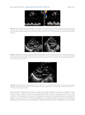
Figure 36. Selected video frames from suprasternal notch view in a neonate with the supra-diaphragmatic type of total anomalous
pulmonary venous connection with pulsed Doppler recording form two different portions (A, B) of the vertical vein (VV); the Doppler
velocities are low, suggesting that there is no obstruction. Reproduced from Ref. [24] . PW: Pulse width; SV: sample volume; WF: width
frequency.
Figure 37. Selected video frames from parasternal long (A) and short (B) axis views showing the truncus (Tr) overriding the ventricular
septum and ventricular septal defect (VSD) (arrows). The position of the VSD (arrow) in the conal septum is demonstrated in (B). In
(A), the left ventricle (LV) is posterior while the right ventricle (RV) is anterior. In (B), the left atrium (LA) is posterior and the RV is
anterior. Reproduced from Ref. [28] .
Figure 38. A parasternal long-axis view showing a truncus arteriosus (TA), a ventricular septal defect (VSD) (arrow) and a truncal
valve overriding the ventricular septum. The VSD is large and measures approximately 8 mm (insert, Dist). Reproduced from Ref. [30] .
LV: Left ventricle; RV: right ventricle.
Pulsed and CW Doppler flow velocities across the branch PAs should be recorded in an attempt to detect
stenosis of these vessels. Detection of associated abnormalities, namely, interruption of the aortic arch,
variations of the origination of the aortic arch branches, coronary artery origin abnormalities, persistent left
superior vena cava, and other VSDs is also feasible by echo-Doppler studies. The presence of narrowing or
insufficiency of the truncal valve and branch PA obstructive lesions may also be identified by echo-Doppler